
In the wake of a car accident, it is a good idea to visit a physician as soon as possible. Getting an MRI After a Car Accident in Brooklyn, NY

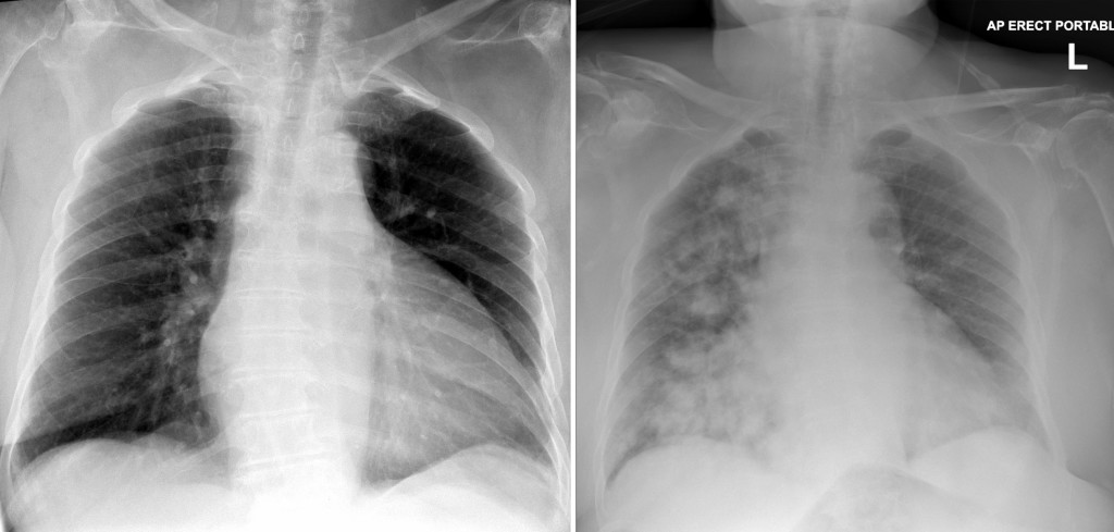
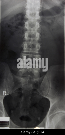
However, the radiation during X-ray imaging is relatively small and roughly the amount you naturally receive over a 10-day period due to background exposure. When getting an X-ray, patients are usually required to protect the other parts of their body from the radiation by wearing a lead apron. Physicians can also look at X-rays of the chest to determine whether a person suffers from pneumonia or other respiratory illness. Mammograms, which take advantage of X-rays, can identify early stages of breast cancer. The rays more easily penetrate soft tissues and fat, so those appear either gray or black.ĭoctors typically use X-rays to check for broken bones (fractures), but they also have other medical purposes. The calcium contained in bones means they absorb the most radiation, so they appear white on the X-ray film. The images are black and white because various tissues absorb varying amounts of radiation. Imaging utilizing this type of radiation has the capability of showing medical professionals the inside of a patient’s body in shades of black and white. X-rays use electromagnetic waves that can penetrate some materials. If you have sustained an injury during a car accident, your physician may order an MRI scan to look for internal injuries. Signs of breast cancer in women and uterine anomalies.Cysts, tumors, and other types of anomalies in various parts of the body.Diseases of the liver and other abdominal organs.Abnormalities or injuries to the joints.Medical professionals commonly use an MRI scanner to view or look for: Physicians can perform an MRI on various parts of the body, but it is especially useful for viewing the nervous system and soft tissues. Unlike a computed tomography (CT) scan or X-ray, MRIs don’t involve ionizing radiation. Physicians often use this type of test to diagnose patients and to see how well they have responded to treatment.

Magnetic resonance imaging, more commonly referred to as MRI, is a medical imaging test that utilizes radio waves, strong magnets, and a computer to create detailed images of the inside of the body. X-ray, there are many factors to think about. There are key differences between these two types of testing, and which you undergo depends largely on the nature of your injury and your physician’s choice. Two types of diagnostic imaging, magnetic resonance imaging (MRI) and X-ray, are common radiology services for car accident survivors. Fortunately, diagnostic imaging can help physicians and other medical professionals detect such injuries and come up with a proper treatment plan for you. Getting a proper diagnosis as soon as possible is your top priority. As an example, we often use computer-generated 3D modeling (based on CT scans) to complete preoperative planning for knee and hip replacement surgeries.A car accident can leave you with painful and lasting injuries or medical conditions. 3D modeling is accomplished with sophisticated computer programs and based on actual patient imaging (xray, CT, and/or MRI). Increasingly 3D modeling is used to provide surgeons with a precise 3D image of bone and joint structures. MRI scans typically take longer and are more expensive than xrays and CT scans. MRI machines use magnetic fields and computer processing to take high-resolution pictures of bones and soft tissues.ĭue to the ability to generate more detailed images – especially of soft tissues - MRI can be used to better evaluate ligaments, tendons and cartilage surfaces. Unlike xrays and CT scans, MRI does not require the use of radiation. Magnetic resonance imaging (MRI) uses radio waves to produce detailed cross-sectional images of the body. CT scans are more expensive and take more time than regular xray imaging. CT scans allow surgeons to visualize 3D size, shape, and position of structures that are deeper within the body and joints. Computed tomography (CT)ĬT is an imaging technique that combines xray images from various angles with computer technology that produces more detailed, cross-sectional images of the body. Less dense soft tissues structures allow the radiation pass-through, therefore appear darker on xray films. More dense objects appear white or lighter on xray films because they absorb the radiation. Xrays use low doses of radiation to image dense objects including bones, calcifications, some tumors, and other dense matter. Even if a more complex test is required for final diagnosis, xray is often used as a first-line screen. Due to cost and availability, xrays (radiographs) are the most commonly ordered diagnostic imaging modality.


 0 kommentar(er)
